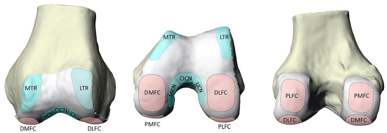
Figure 2.
A left distal femur model demonstrating 5 donor locations (blue): the lateral trochlear ridge (LTR), medial trochlear ridge (MTR), lateral intercondylar notch (LICN), and medial intercondylar notch (MICN) and 4 recipient (pink) regions: the distal medial femoral condyle (DMFC), distal lateral femoral condyle (DLFC), posterior medial femoral condyle (PMFC), and posterior lateral femoral condyle (PLFC). (A) Anterior view which best visualizes the trochlear donor regions. (B) Distal view which best visualizes the distal femoral condyles recipient regions and the intercondylar notch donor regions. (C) Posterior view which best visualizes the posterior condyles recipient regions.

