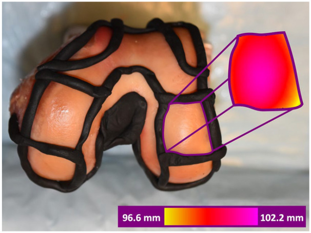
Figure 3.

Top: Distal view of a cadaveric left distal femur with sectioned regions. Plastalina Modeling Clay (Van Aken International, Dalton, GA) bounds the segmented regions. Right colored section: Corresponding scanned images of the distal lateral femur condyle captured with the Gocator scanner. Coloring denotes the distance between the scanner camera and the articular surface.
