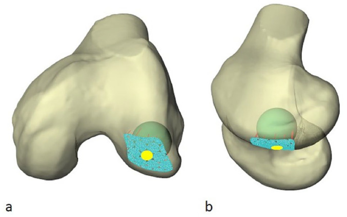
Figure 4.

Two views (a, semiaxial; b, semisagittal) of a left distal femur illustrating a “best fit” radius of curvature (RoC) sphere within the distal lateral femoral condyle. The honeycomb pattern of juxtaposed circles is formed by blue nodes. The contacted articular surface highlighted in yellow represents one simulated graft recipient site. The peripheral partial blue circles were excluded.
