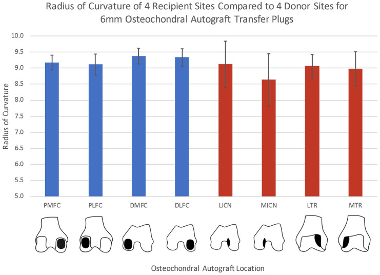
Figure 5.
Bar graph with average radius of curvature (RoC) of 4 recipient locations (blue) and 4 donor locations (red) for 6-mm diameter osteochondral autograft transfers. Donor regions included the lateral trochlear ridge (LTR) and medial trochlear ridge (MTR), as well as the lateral intercondylar notch (LICN) and medial intercondylar notch (MICN). Recipient regions analyzed were the distal medial (DMFC), posterior medial (PMFC), distal lateral (DLFC), and posterior lateral femur condyle (PLFC).

