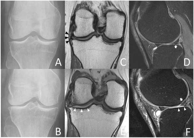
Figure 2.
Index visit (A) and 4-year follow-up radiographs (B) of a 69-year-old female accelerate knee osteoarthritis (AKOA) subject show Kellgren-Lawrence (KL) grade progression from KL 0 to KL 4 of the right knee within 4 years. Index visit coronal (C) 3-T magnetic resonance (MR) images depict complex tearing of the lateral meniscus body and extrusion of 4 mm (black arrowheads) and index visit sagittal (D) MR images show a complex tear of the lateral anterior horn (white arrow). At 4-year follow-up (E and F) the lateral meniscus body is extruded by 5 mm (black arrowheads) and there is a small meniscal cyst at the anterior horn (white arrow). Note full-thickness cartilage loss of the lateral tibia and femur (white arrowheads), joint effusion, and loose bodies in the joint (black arrow).

