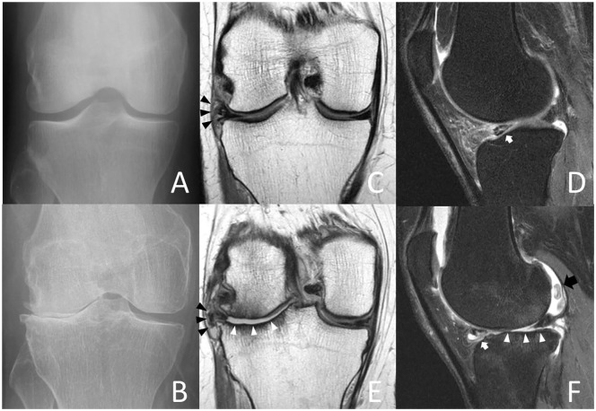
Figure 3.
Index visit (A) and 4-year follow-up radiographs (B) of a 53-year-old female accelerated knee osteoarthritis (AKOA) subject with Kellgren-Lawrence (KL) grade progression from KL 1 to 3 of the right knee within 4 years. Coronal (C) and sagittal (D) index visit 3-T magnetic resonance (MR) images show intrasubstance degeneration of the lateral meniscus body with a tear extending to the interior surface and extrusion (3 mm, black arrowheads). Note irregularity, fraying, and abnormal signal of the lateral posterior horn (white arrow) and diffuse thinning of the lateral tibial plateau cartilage. At 4-year follow-up (E and F) extensive cartilage loss of the lateral femur and tibia (white arrowheads), increased degeneration of the lateral posterior horn with tearing (white arrow) and increased intrasubstance degeneration of the anterior horn with a large meniscal cyst (F).

