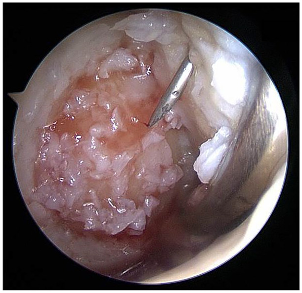
Figure 5.

Arthroscopically prepared cartilage defect (medial femoral condyle, right knee joint, dry conditions; defect from Fig. 2 ). The cartilage chips (previously mixed with platelet-rich plasma [PRP]) have been implanted already via arthroscopic techniques. An autologous thrombin solution is currently applied via a needle through the proximal portal for final fixation of the chips. As the last step, a mix of PRP and the autologous thrombin solution is applied to finalize the coagulation cascade in order to provide initial construct stability. In the distal portal, a swab is placed in order to safely keep the joint dry and to tense the joint capsule and Hoffa fat to distance them from the defect.
