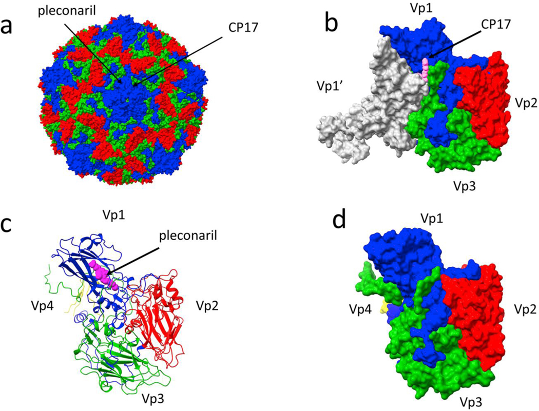
Figure 1. Antiviral binding sites on a picornavirus capsid.
(a) A coxsackie virus capsid (1cov), with the viral structural proteins Vp1–3 colored blue, red, and green, respectively; Vp4 is inside the capsid and not visible in this image. Two distinct sites for capsid-directed antiviral compounds are denoted with arrows. (b) The binding site for compound 17 (CP17, pink), from Abdelnabi et al. 2019 (6gzv), is at an interface of a Vp3 and the two adjacent Vp1 molecules (one shown in white). The view is from the outside of the capsid. (c) In contrast, the binding location for pleconaril (magenta), binding the same site as a WIN compound, only contacts a single copy of Vp1 and is not located at an intermolecular interface (1ncr). (d) A surface representation of the same complex as (c) emphasizes that pleconaril is completely enclosed by Vp1.

