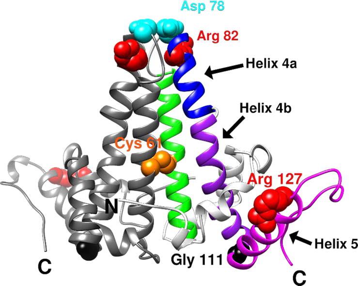
Figure 1.
Ribbon representation of an HBV Cp149 dimer (PDB: 1QGT) and key residues discussed in this study. One half-dimer is shown in gray and the other is divided into different regions by colors: helix 4 is divided into helix 4a (blue) and 4b (purple). The interdimer interface is formed by interactions between helix 5 (magenta) from one dimer and the loop at the end of helix 5 from a neighboring dimer. Key residues are shown in space-filled representation: Cys61 in orange, Asp78 in cyan, Gly111 in black, Arg82 and Arg127 in red.

