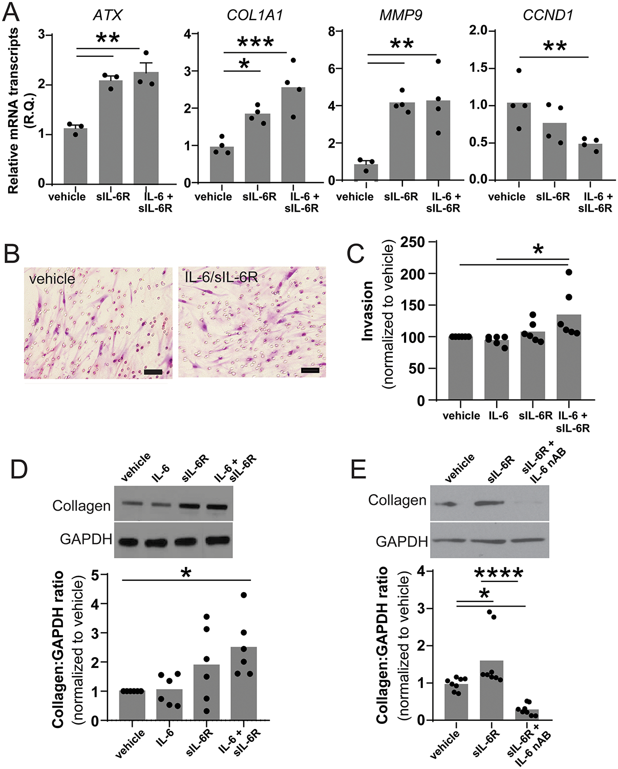
Figure 3. IL-6 trans-signaling stimulates cellular invasion and fibrotic differentiation.

(A) mRNA was harvested from MCs treated with either vehicle or the combination of IL-6 and sIL-6R for 24 hours. Expression of known STAT3-regulated genes was assessed using qPCR (n=4, * p<0.05 ** p<0.01 *** p<0.001). (B) The invasiveness of MC treated with vehicle, IL-6, sIL-6R or IL-6/sIL-6R was measured using a standard matrigel transwell invasion assay and qualitatively assessed by cytology and quantitatively evaluated by MTT assay. Representative image of MC which migrated through the matrigel to the underside of the transwell membrane. (scale bar represents 50 μm). (C) The combination of IL-6/sIL-6R caused a 37% increase in invasion compared to vehicle control (100.0±25.9 versus 136.9±23.14, n=6, p<0.05) (D) Co-treatment with IL-6 and sIL-6R stimulated significant increase in collagen protein expression (1.0 vs 2.492±1.03, n=6, *p<0.05). (E) sIL-6R-induced collagen expression was completed blocked by the addition of an IL-6 neutralizing antibody (1.00±0.18 vs 1.642±0.85 vs 0.295±0.17, n=4, *p<0.05 ****p<0.0001).
