Figure 6.

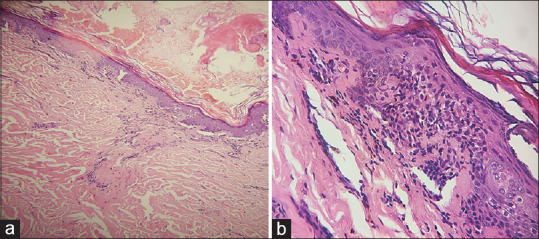
(a) Skin biopsy shows hyperkeratosis, mild atrophy, perivascular and periadnexal chronic inflammatory infiltrate (H and E, ×100). (b) Prominent vacuolization of basal cell layer of epidermis (H and E, ×400)

(a) Skin biopsy shows hyperkeratosis, mild atrophy, perivascular and periadnexal chronic inflammatory infiltrate (H and E, ×100). (b) Prominent vacuolization of basal cell layer of epidermis (H and E, ×400)