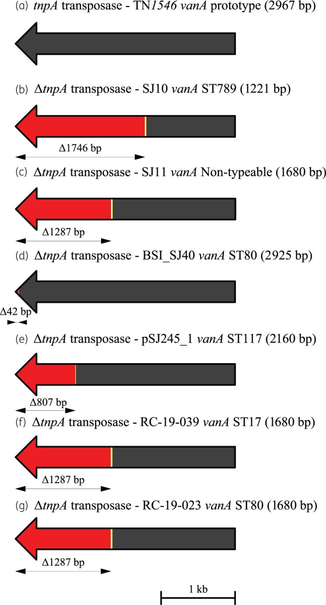
Figure 4.

Schematic diagram showing the extent of deletions and other mutations in the tnpA transposase gene within the vanA region closed by hybrid assembly from VREfm isolates from Irish hospitals. The arrows indicate the direction of transcription. Deletions are shown in red, bp substitutions are indicated in yellow and bp insertions are indicated in green. The extent of sequences deleted (Δ) is shown beneath each tnpA gene in bp. (a) WT tnpA (transposase) gene (2967 bp) from the prototype vanA transposon Tn1546;29 (b) ΔtnpA gene from isolate SJ10 (SJ10vanA, 1221 bp); (c) ΔtnpA gene from isolate SJ11 (SJ11vanA, 1680 bp); (d) ΔtnpA gene from isolate BSI_SJ40 (BSI_SJ40vanA, 2925 bp); (e) ΔtnpA gene from plasmid pSJ245vanA in isolate SJ245 (pSJ245vanA, 2160 bp); (f) ΔtnpA gene from isolate RC_19_039 (RC_19_039vanA, 1680 bp); and (g) ΔtnpA gene from isolate RC_19_023 (RC_19_023vanA, 1680 bp). VREfm isolates were recovered from H1 (b–e), H3 (f) and H4 (g). A reference size scale bar is shown at the bottom of the diagram.
