Clinical Images in Nephrology and Dialysis
Case Description
A 58-year old patient was referred to the kidney-stone clinic when unilateral kidney stones were detected after an abdominal ultrasound to evaluate abdominal pain. Previous urologic evaluation decided against removal of the stones by endourologic procedures. The patient denied any previous history of urinary symptoms, specifically urinary tract infections and gross hematuria, and never had a renal colic or passage of kidney stones. She had no history of gout, inflammatory bowel disease, chronic diarrhea, hyperparathyroidism, neck irradiation, or granulomatous disease, and denied consumption of any lithogenic medication. Family history was negative for kidney stones and she had a healthy identical twin.
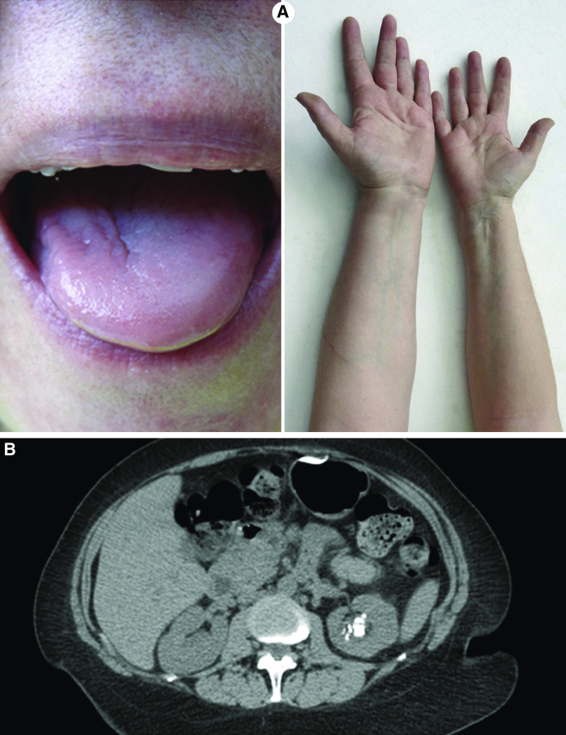
Physical examination was remarkable for a clear asymmetry in the size of the tongue, hands, and legs (Figure 1A). No visceromegaly was detected.
Figure 1.
Hemihypertrophy associated to unilateral nephrocalcinosis. (A) Hemihypertrophy of the left side of the tongue and left arm. (B) Multiple calcifications compatible with nephrocalcinosis in the kidney of the larger body side, with otherwise preserved renal architecture.
Computed tomography scans of the abdomen demonstrated multiple calcifications compatible with nephrocalcinosis in the kidney of the larger body side, with otherwise preserved renal architecture and no evidence of renal cortical scaring. There was no evidence of hydronephrosis (Figure 1B).
The metabolic evaluation revealed adequate urine volume (3.2 L), hypercalciuria (296 mg/24 h), hyperuricosuria (835 mg/24 h), normal urinary citrate (650 mg/24 h), and oxalate excretion (25 mg/24 h). The fasting urinary pH was 5.5, and there was no evidence of renal tubular acidosis. Serum calcium, phosphorus, creatinine, vitamin D metabolites, and parathyroid-hormone levels were normal.
Treatment was initiated with a thiazide diuretic, allopurinol, maintenance of high fluid intake, and a low-sodium diet.
The patient showed evidence of congenital hemihypertrophy, a condition characterized by asymmetry of the body as a result of hypertrophy of all somatic elements (muscles, bones, nerves, vessels) of one or more parts of the body. Congenital hemihypertrophy has been estimated to occur in 1:40,000 live births and has been associated with other developmental abnormalities, including Beckwith–Wiedemann syndrome—a growth disorder characterized by macrosomia, macroglossia, visceromegaly, embryonal tumors (Wilms tumor, hepatoblastoma, neuroblastoma, and rhabdomyosarcoma), omphalocele, poly- and syndactyly, facial nevus, earlobe creases and pits, and multiple renal abnormalities (1). Beckwith–Wiedemann syndrome is a model disorder for the study of imprinting, growth dysregulation, and tumorigenesis. The most common molecular cause is hypomethylation of the maternal imprinting control region 2 (ICR2) in 11p15 DNA, and methylation tests of 11p15.
Renal abnormalities observed in Beckwith–Wiedemann syndrome include medullary dysplasia, nephrocalcinosis, medullary sponge kidney, and nephromegaly. Up to 10% of patients with congenital hemihypertrophy have medullary sponge kidney, a condition frequently associated with urolithiasis (2). The cause for the unilateral nephrocalcinosis ipsilateral to the hemihypertrophic side remains unclear. A study evaluating laterality of nephrocalcinosis in other conditions, such as severe hypocitraturia, suggest that additional intrinsic local factors such as renal perfusion or vascular injury must also play a role in stone formation (3).
Teaching Points
Congenital hemihypertrophy (Beckwith–Wiedemann syndrome) is characterized by asymmetry of the body as a result of hypertrophy of all somatic elements (muscles, bones, nerves, vessels) of one or more body parts.
Renal abnormalities observed in Beckwith–Wiedemann syndrome include medullary dysplasia, nephrocalcinosis, medullary sponge kidney, and nephromegaly. Up to 10% of patients with congenital hemihypertrophy have medullary sponge kidney, a condition frequently associated with urolithiasis.
A large proportion of patients can develop, for unknown reasons, unilateral nephrocalcinosis.
Disclosures
Dr. Weisinger and Dr. Freundlich have nothing to disclose.
Funding
None.
Author Contributions
J.R. Weisinger and M. Freundlich reviewed and edited the manuscript.
References
- 1.Weksberg R, Shuman C, Beckwith JB: Beckwith-Wiedemann syndrome. Eur J Hum Genet 18: 8–14, 2010 [DOI] [PMC free article] [PubMed] [Google Scholar]
- 2.Indridason OS, Thomas L, Berkoben M: Medullary sponge kidney associated with congenital hemihypertrophy. J Am Soc Nephrol 7: 1123–1130, 1996 [DOI] [PubMed] [Google Scholar]
- 3.Le JD, Eisner BH, Tseng TY, Chi T, Stoller ML: Laterality of nephrocalcinosis in kidney stone formers with severe hypocitraturia. BJU Int 107: 106–110, 2011 [DOI] [PMC free article] [PubMed] [Google Scholar]


