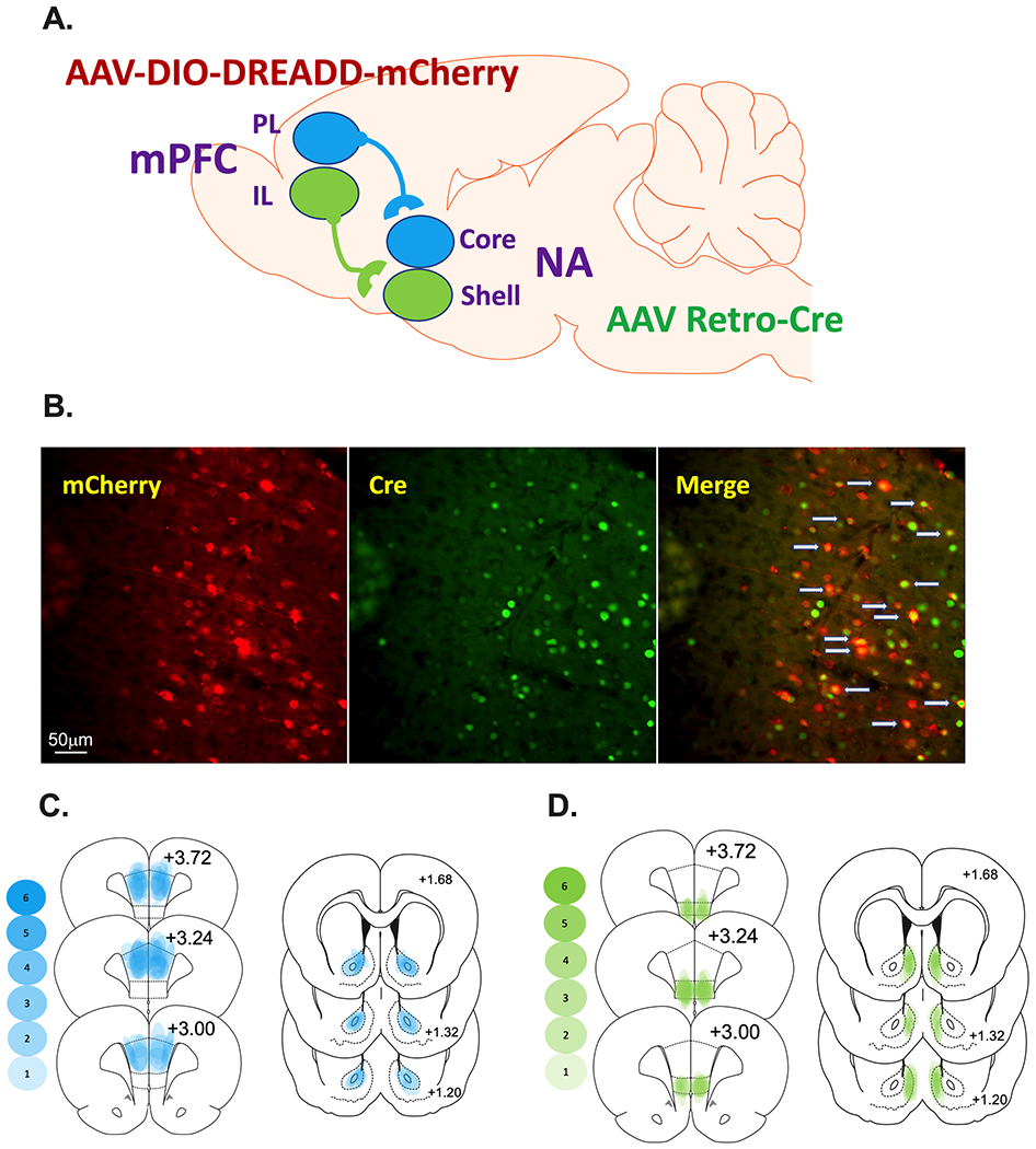
Figure 1.

Schematic representation of the prelimbic (PL)-nucleus accumbens (NA)core and infralimbic (IL)-NAshell subcircuits as well as the dual viral approach. A. AAVrg-pmSyn1-enhanced blue fluorescent protein(eBFP)-Cre or AAVrg-hSyn-HI-enhanced green fluorescent protein(eGFP)-Cre was microinjected into the NAcore or NAshell depending on the experiment. The Cre-dependent Gi-DREADD [AAV-hSyn-DIO-hM4D(Gi)-mCherry or Gq-DREADD [AAV-hSyn-DIO-hM3D(Gq)-mCherry] into the PL or IL (i.e., the cell body area) depending on the experiment. As a control, some rats received DIO-mCherry [AAV-hSyn-DIO-mCherry] instead of DREADDs into the cortical area under similar parameters. B. (Left panel) IHC detection of DIO-hM4Di-mCherry (red) in the PL. (Center panel) IHC detection of GFP (green) in the PL. (Left panel). Overlay of the cells expressing Cre merged with those expressing mCherry are identified with gray arrows (Right panel). C and D. Heat maps were generated for the PL-NAcore (C) and IL-NAshell (D) to represent the extent of viral transduction in each region. Transparent blue or green shapes were used to yield more saturation when viral placements were consistent across animals.
