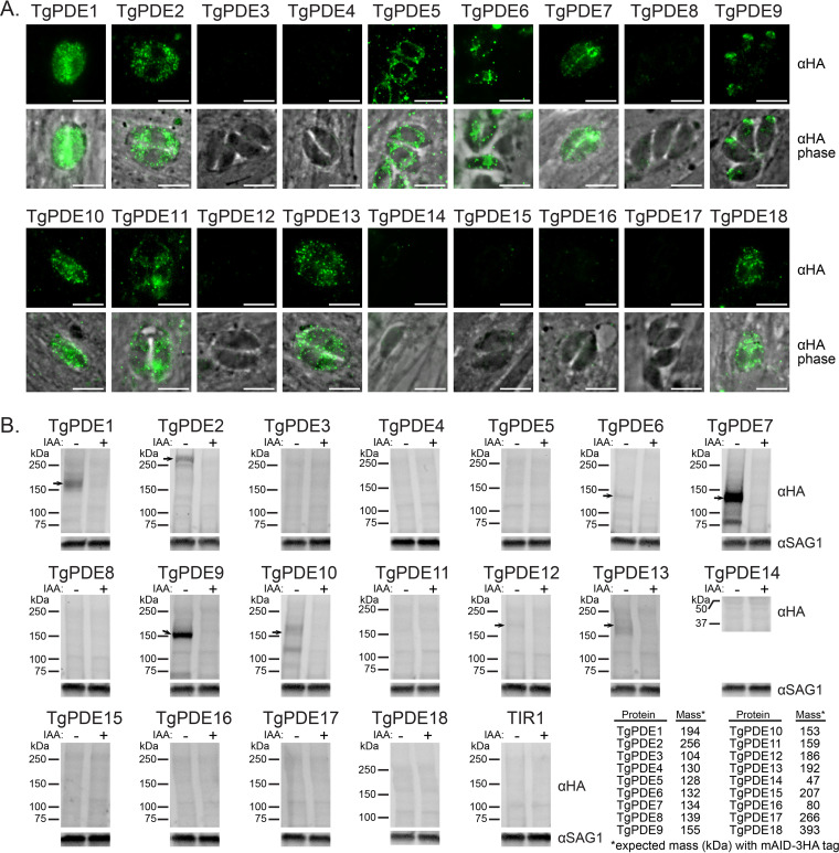
FIG 3.
Expression and depletion of TgPDE-mAID-3HA fusions in tachyzoites. (A) IF microscopy of intracellular RH TgPDE-mAID-3HA parasites labeled with mouse anti-HA and goat anti-mouse IgG Alexa Fluor 488. Bar, 5 μm. (B) Immunoblots of lysates from RH TIR1-3FLAG (TIR1) and RH TgPDE-mAID-3HA (TgPDE) parasites treated with vehicle (EtOH) or 0.5 mM IAA for 18 h. Blots were probed with mouse anti-HA, rabbit anti-SAG1, goat anti-mouse-AFP800, and goat anti-rabbit-AFP680. The table presents the predicted total mass of each TgPDE, including the mAID-3HA tag (12 kDa). Arrows indicate immunoblot-detectable TgPDE-mAID-3HA fusions in tachyzoites.

