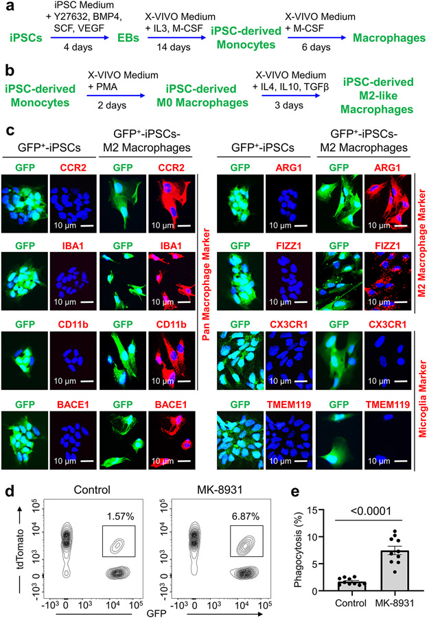
Extended Data Fig. 1. Derivation of Macrophages from Human iPS Cells (iPSCs).
a, A brief protocol for generating monocytes and macrophages from human iPS cells (iPSCs) expressing GFP. EB: Embryonic body; PMA: Phorbol 12-myristate 13-acetate.
b, A brief protocol for the generation of M2-like macrophages (iPSC-M2) from iPSC-derived monocytes expressing GFP.
c, Immunofluorescent analysis of the pan macrophage markers (CCR2, CD11b and IBA1; in red), the M2 macrophage makers (ARG1 and FIZZ1; in red), BACE1 (in red), or the microglia-specific markers (TMEM119 and CX3CR1; in red) in iPSCs-M2 macrophages. Representative immunofluorescent images show expression of BACE1, the pan macrophage markers, and the M2 macrophage markers in iPSC-M2 macrophages.
d,e, Flow cytometric analyses of macrophage phagocytosis of human glioma cells in vitro. Representative flow cytometric plots (d) shows the gating strategy for identifying macrophage phagocytosis of glioma cells (CCF-3264, tdTomato+) in iPSC-derived macrophages (GFP+) that were pre-treated with MK-8931 (50 μg/mL) or vehicle control for 48 hours. Quantification (e) of the flow cytometric analyses shows fractions of macrophage phagocytosis of glioma cells (GFP+/tdTomato+). Data are shown as means ± SEM. n = 10 independent phagocytosis assays per group. Statistical significance was determined by two-tailed Student’s t-test; p<0.0001.

