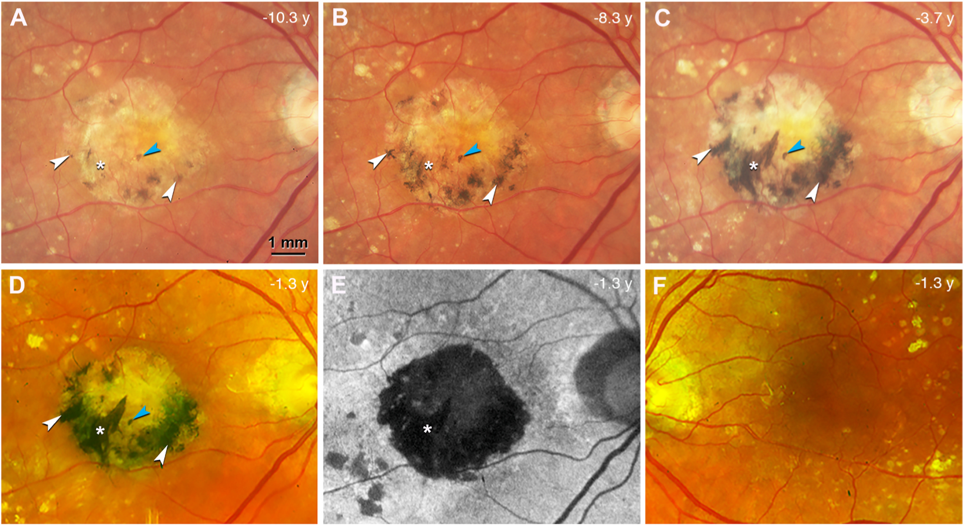
Fig. 2. Black pigment evolution in the index eye with neovascular age-related macular degeneration during 9 years of follow-up.

A-D. Color fundus photograph (CFP) of the right eye, acquired at different time points before patient death (in years) as indicated, shows a central subretinal fibrosis with increasing and darkening black pigment (white arrowheads). A prominent spear-like streak of black pigment developed over this time (white asterisk). One small spot of black pigment maintains stable during 9 years of follow up (blue arrowhead). Topcon camera, A-C; Optos California (Dunfermline Scotland), D. E. Fundus autofluorescence (FAF) at the last clinical visit, the same time point as in (D), shows that the fibrotic region is hypoFAF. The spear-like streak with black pigment is deeply hypoFAF (white asterisk). F. CFP of the left eye at the last clinical visit shows no black pigment or subretinal fibrosis.
