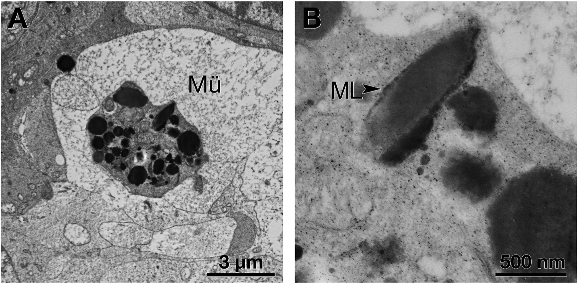
Fig. 6. Cellular fragment containing retinal pigment epithelium (RPE) organelles within Müller glia.

A. A Müller glial (Mü) process has enveloped an RPE cell fragment, recognized by its characteristic organelles. B. At higher magnification, an elongated organelle resembles that labeled melanolipofuscin (ML, black arrowhead) (Feeney, 1978).
