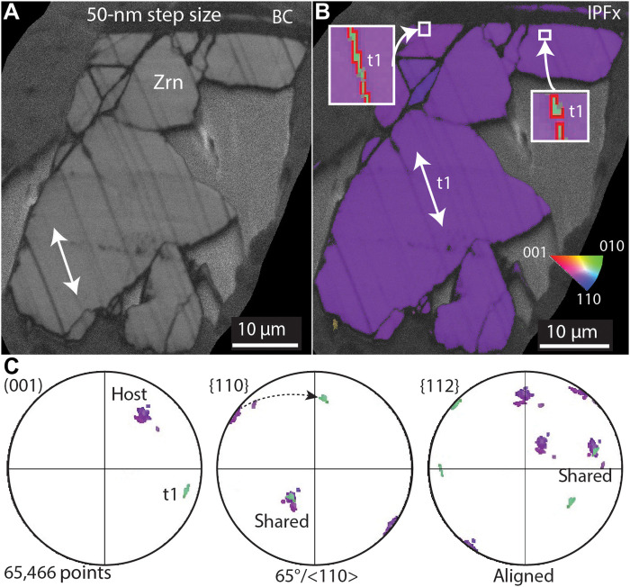
Fig. 2. Scanning electron microscope images of a shock-deformed zircon grain from NWA 7034.
(A) Band contrast (BC) map of zircon (Zrn) grain showing planar microstructures. (B) Inverse pole figure (IPFx) showing one orientation of {112} twins. (C) Stereo plot showing the orientation relationship of the host grain and {112} shock deformation twins. Stereonets are equal area, lower hemisphere projections in the sample reference frame. t1, zircon twin.

