Figure 3.
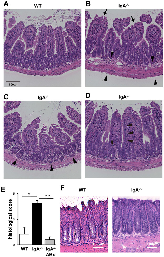
Pathological changes in the ileum tissues of IgA−/−. Tissues of WT (A) and IgA−/− (B–D) were fixed and stained with H&E. Normal appearance at ileum of WT (A), mild infiltration with mononuclear cells in the lamina propria (arrows in B), thickened submucosal, muscular and serosal layers (arrowheads in B), crypt abscesses (arrowheads in C), reduced goblet cells (arrowheads in D) at ileum of IgA−/− are indicated. Representative results are shown (n>7). (E) Pathological evaluation of the ileum. WT (left), IgA−/− (middle) and antibiotic (ABx)-treated IgA−/− (right) mice were subjected for the analysis. Scores were determined as described in online supplemental materials and methods. Values are presented as means±SEM. *p<0.05. **p<0.01. (F) Histological evaluation of the colons from WT (left) and IgA−/− (right). Representative results are shown (n>7).

