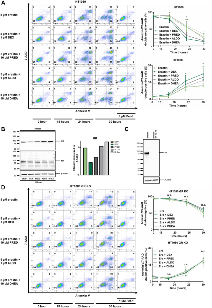
Fig. 2. Steroid-induced sensitization to erastin-induced ferroptosis requires the GR 1.
(A) HT1080 cells were treated with erastin, Fer-1, 1 μM dexamethasone (DEX), 10 μM prednisolone (PRED), 1 μM aldosterone (ALDO), and 10 μM dehydroepiandrostendione (DHEA) as indicated. Primary FACS plots and respective quantifications of indicated populations are demonstrated. Note the sensitization to ferroptosis induced by DEX and PRED, but not by ALDO and DHEA. (B) Assessment of GR and mineralocorticoid receptor (MR) down-regulation in response to indicated steroid hormones. β-Actin serves as a loading control. (C) CRISPR-Cas9–mediated knockout of GR from HT1080 cells, confirmed by Western blotting. (D) Experiment performed as described for (A), but using GR-knockout cells. Note the loss of sensitization to ferroptosis. All experiments shown are representative of at least three independent complete repetitions performed. The graphs show means ± SD. Statistical analysis was performed using Student’s t test for each time point. *P ≤ 0.05, **P ≤ 0.01, n.s., nonsignificant.

