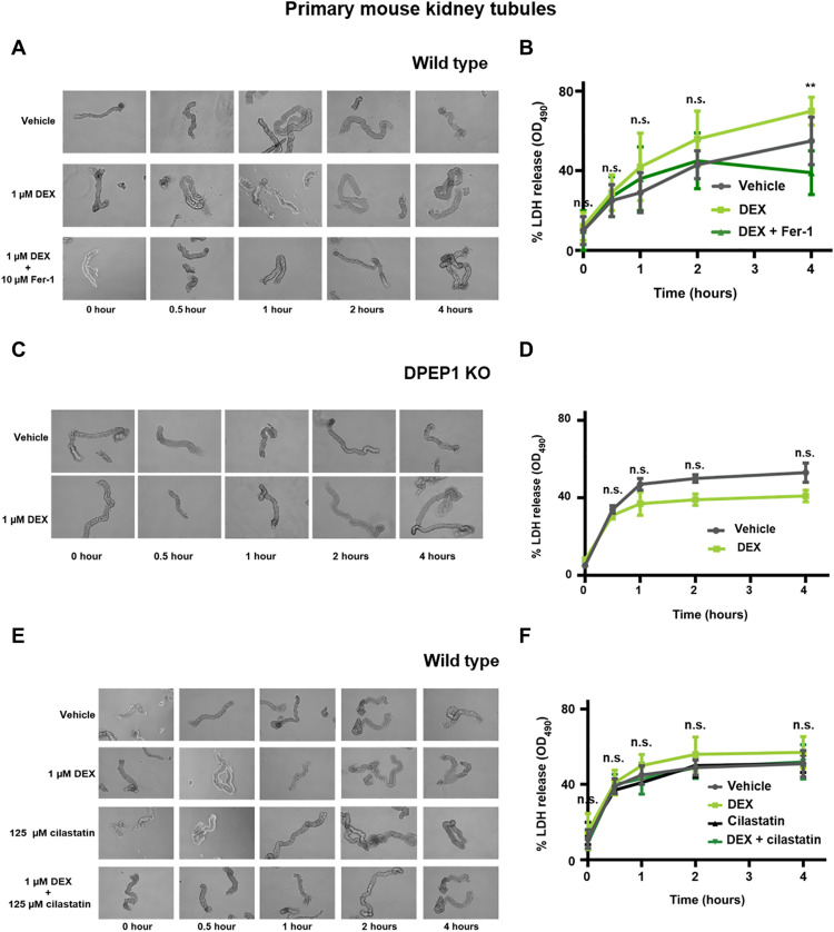
Fig. 5. Ferroptosis in freshly isolated renal tubules is accelerated by dexamethasone in wild-type but not in DPEP1 knockout mice.
(A) Representative images of freshly isolated murine kidney tubules undergoing spontaneous cell death in the presence of either vehicle or 1 μM dexamethasone in the presence or absence of 10 μM Fer-1. (B) LDH release of respective time points. (C) Representative images of freshly isolated murine kidney tubules from DPEP1 knockout mice undergoing spontaneous cell death in the presence of vehicle or 1 μM dexamethasone. (D) LDH release of respective time points. (E) Representative images of freshly isolated murine kidney tubules undergoing spontaneous cell death in the presence of either vehicle, 1 μM dexamethasone, 125 μm cilastatin, or dexamethasone and cilastatin. (F) LDH release of respective time points. The graphs show means ± SD. Statistical analysis was performed using Student’s t test for each time point. **P ≤ 0.01.

