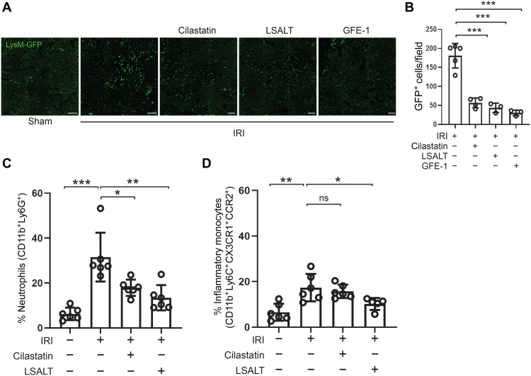
Fig. 4. DPEP1 inhibitors and leukocyte recruitment during kidney IRI.
(A) Kidney IVM in LysMgfp/gfp mice at 2 hours following IRI in mice treated with LSALT peptide, cilastatin, or GFE-1 peptide. Sham-operated kidney is used as a negative control. Scale bars, 100 μm. (B) Stationary GFP+ leukocytes/field in the kidney were quantified (versus IRI: cilastatin, ***P = 0.0002; LSALT, ***P = 0.0005; GFE-1, ***P = 0.0003; n = 3 to 5 per group, ANOVA with Dunnett’s post hoc test). (C and D) Flow cytometry of leukocytes isolated from ischemic (IRI) and contralateral (Cntrl) kidneys in wild-type mice treated with or without cilastatin or LSALT peptide at 48 hours (neutrophils: Cntrl versus IRI, ***P = 0.0006; IRI versus IRI + cilastatin, *P = 0.011; IRI versus IRI + LSALT, P = 0.001; n = 5 to 6 per group, ANOVA with Dunnett’s post hoc test) (inflammatory monocytes: Cntrl versus IRI, **P = 0.01; IRI versus IRI + cilastatin, ns = not significant; IRI versus IRI + LSAL, P = 0.047; n = 5 to 6 per group, ANOVA with Dunnett’s post hoc test).

