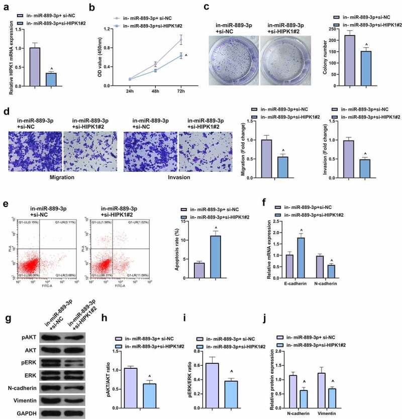
Figure 5.

HIPK1 overexpression motivates the growth and metastasis of LC cells
A. qPCR to detect the expression of HIPK1 after elevation of HIPK1; B/C. CCK-8 and plate cloning to detect the proliferation of cells after elevated HIPK1; D. Transwell to detect the invasion and migration abilities of cells after elevated HIPK1; E. Cell apoptosis after elevated HIPK1 detected by flow cytometry; F. EMT-related molecules E-cadherin and N-cadherin detected by PCR; G. Western Blot analysis of pAkt, Perk, N-cadherin and Vimentin protein levels. The data in the figures were all measurement data in the form of mean ± SD. & vs the oe-NC, P < 0.05.
