
Figure 2.
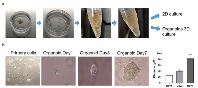
Images representing the process of culturing organoids from an adenomyoepithelioma (AME) of a patient. (a) The AME tumor sample was cut into 1-mm3 pieces and digested with 2 mg/mL collagenase. The tumor cells were then cultured under 2D and 3D culture systems. (b) The morphology of the cultured tumor cells and organoids at days 1, 3, and 7, as observed under a microscope. The diameter of the organoids was measured at days 1, 3, and 7. Data are presented as mean ± standard deviation. n = 3, *p < 0.05, ***p < 0.001