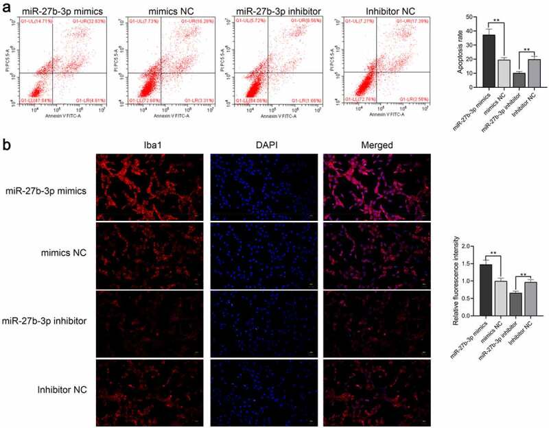
Figure 4.

miR-27b-3p accelerated apoptosis and Iba1 expression inmicroglial cells treated with 100 ng/mL LPS (n = 3). (a) Flow cytometry for detecting apoptosis of miR-27b-3p in microglia. (b) Immunofluorescence for detecting Iba1 expression. **, P < 0.01 compared with the NC control
