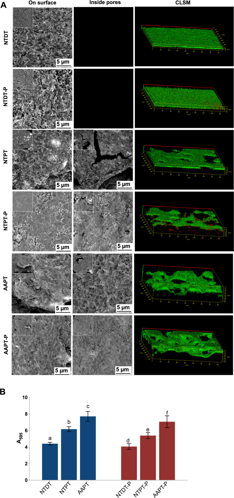
Fig. 4.

S. mutans adhesion was finally observed at the endpoint (24 h) of incubation on Ti with or without serum precoating. A SEM and CLSM images showed the morphology of bacterial adhesion on NTDT, NTDT-P, NTPT, NTPT-P, AAPT, and AAPT-P. The results showed that more S. mutans cells clustered to form biofilms coating all sample surfaces. Bacterial aggregation on AAPT and AAPT-P seemed to be more evident than on NTPT and NTDT as well as on NTPT-P and NTDT-P. B Quantification of bacterial adhesion in each group was performed by measuring the absorbance at 595 nm (n = 4). The results showed that bacteria adhered more to porous samples than to dense samples (p < 0.05). It was also evident that protein coating tended to prevent the bacteria from adhering to material surfaces
