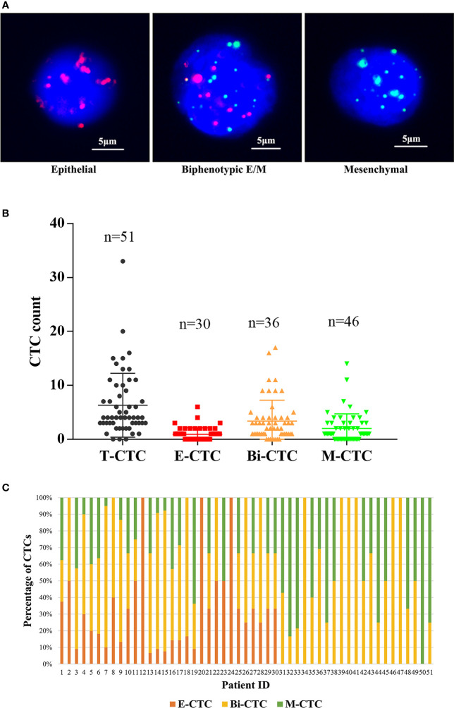
Figure 2.
(A) Representative fluorescence images of three types of CTCs isolated from the peripheral blood of omHSPC patients based on RNA-ISH staining for leukocytes (CD45, white), epithelial cells (EpCAM and CK8/18/19, red), and mesenchymal cells (vimentin and twist, green). 4´,6-Diamidino-2-phenylindole was used to stain cell nuclei (blue). The scale bar indicates 5 μm. (B) Levels of CTC subtypes. (C) The distribution of three subtypes of CTCs in each patient.

