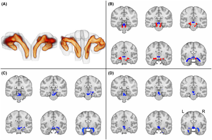
FIGURE 9.

Comparison of GO‐ESP with state‐of‐the‐art approach. A, The 3D view of GO‐ESP generated pathways from the back of brain. Left, Principal EMI generated using our spatially weighted seeding region (gray outline). Right, Principal EMI generated using dilated mask of medial pathway from Sun et al. (2020) as seeding region (grey outline). B, Red, Principal EMI generated from GO‐ESP using LC, amygdala, and dilated thalamus as inclusion regions and VTA as an exclusion region. Blue, Medial track from Sun et al. (2020). C, Replication of medial pathway in MRtrix using seeding approach from Sun et al. (2020). D, Generation of medial pathway in MRtrix seeding from LC and using the TEC as the only inclusion region
