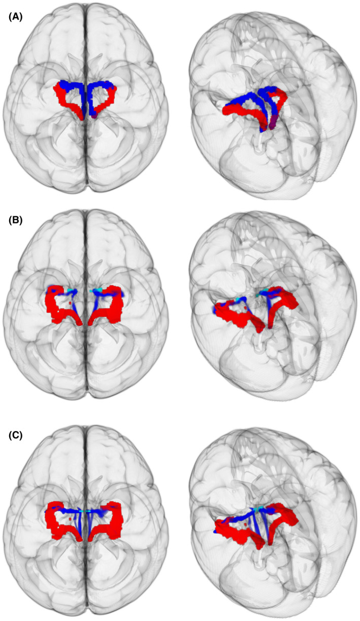
FIGURE 10.

Extracting “medial” pathway using GO‐ESP in HCP subjects. Left, View from top of brain. Right, View from top left angle. A, Extraction of both pathways with widely distributed seeding strategy. Red, Principal EMI. Blue, Third EMI for left hemisphere, and second EMI for the right hemisphere. B, Red, Principal GO‐ESP generated EMI. Blue, Principal GO‐ESP generated EMI using anterior‐medial amygdala (light blue) as additional seeding region. C, Red, Principal GO‐ESP generated EMI. Blue, Principal GO‐ESP generated EMI using a medial disc ROI (light blue) encompassing the anterior‐medial portion of pathway from Sun et al. (2020)
