
Figure 4b.
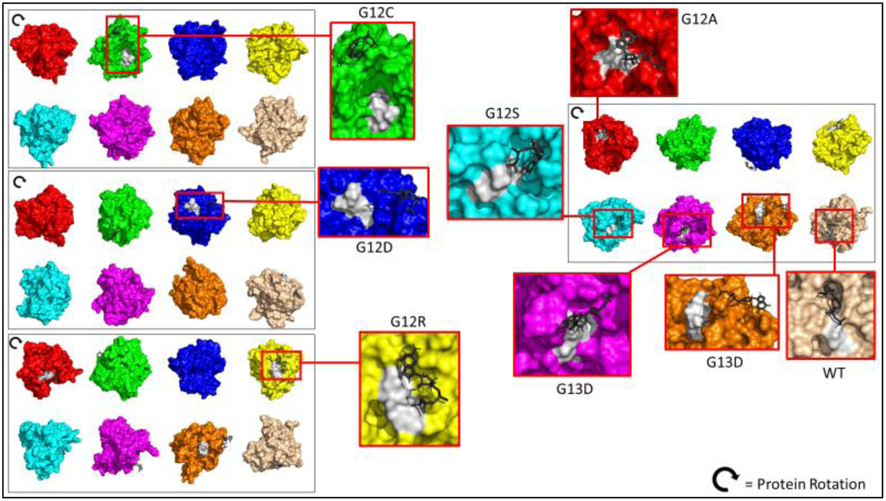
Surface KRAS in silico Protein Mutants and Wildtype 3D structure. PyMOL 3D representation of each KRAS protein bound to their respective substrate (shaded black) in its conformation. Shaded white represents the p-loop region. WT has GTP molecule sitting inside pocket of KRAS protein, while mutants are not in proper conformation.