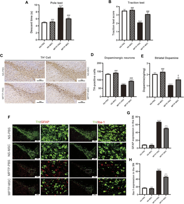
FIGURE 3.
UC-MSCs improved motor function, protected dopaminergic neurons in the substantia nigra and striatum, and alleviated microglia-mediated neuroinflammation in MPTP-induced PD mice. (A) Pole test; (B) traction test; (C) Immunohistostaining for tyrosine hydroxylase (TH) in the SN; (D) quantitative analysis of the number of TH-positive cells in the SN; (E) content of dopamine was measured by HPLC-MS in the ST. Data of (A,B) (n = 12 per group) are expressed as mean ± SE. Data of (C,D) (n = 3–4 per group) are expressed as mean ± SE. Scale bar: 100 μm (SN). (F) Double immunofluorescence staining for TH (green), GFAP (red), and Iba-1 (red) in the SN; (G) Quantitative analysis of the number of GFAP positive cells in each group; (H) Quantitative analysis of the number of microglia in each group; Data of (F–H) (n = 4 per group) are expressed as mean ± SE. Scale bar: 100 μm (SN). *p < 0.05, **p < 0.01, ***p < 0.001 compared with NS-PBS group, ## p < 0.01, ### p < 0.001 compared with MPTP-PBS group by one-way ANOVA.

