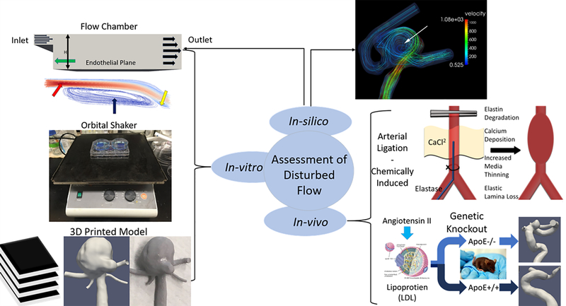
Figure 4:
Overview of methodologies for studying disturbed flow in the vasculature/aneurysms. In-vitro models such as step-down flow chambers (step-down, inlet, disturbed flow, flow re-attachment: green, red, blue, and yellow arrows respectively), orbital shaker, and 3D printed vascular models (medical imaging data, computational model, 3D printed) allow the study of cultured cells under controlled flow. In-vivo techniques such as arterial ligation, chemically induced aneurysms (elastase injection or CaCl2 exposure), and genetic knockout animals (lipoprotein image from https://www.britannica.com/science/lipoprotein#/media/1/342894/92254). In-silico simulated flow is often used alongside other methodologies to analyze flow characteristics within 3D in-vitro models or in-vivo developed aneurysms (white arrow).

