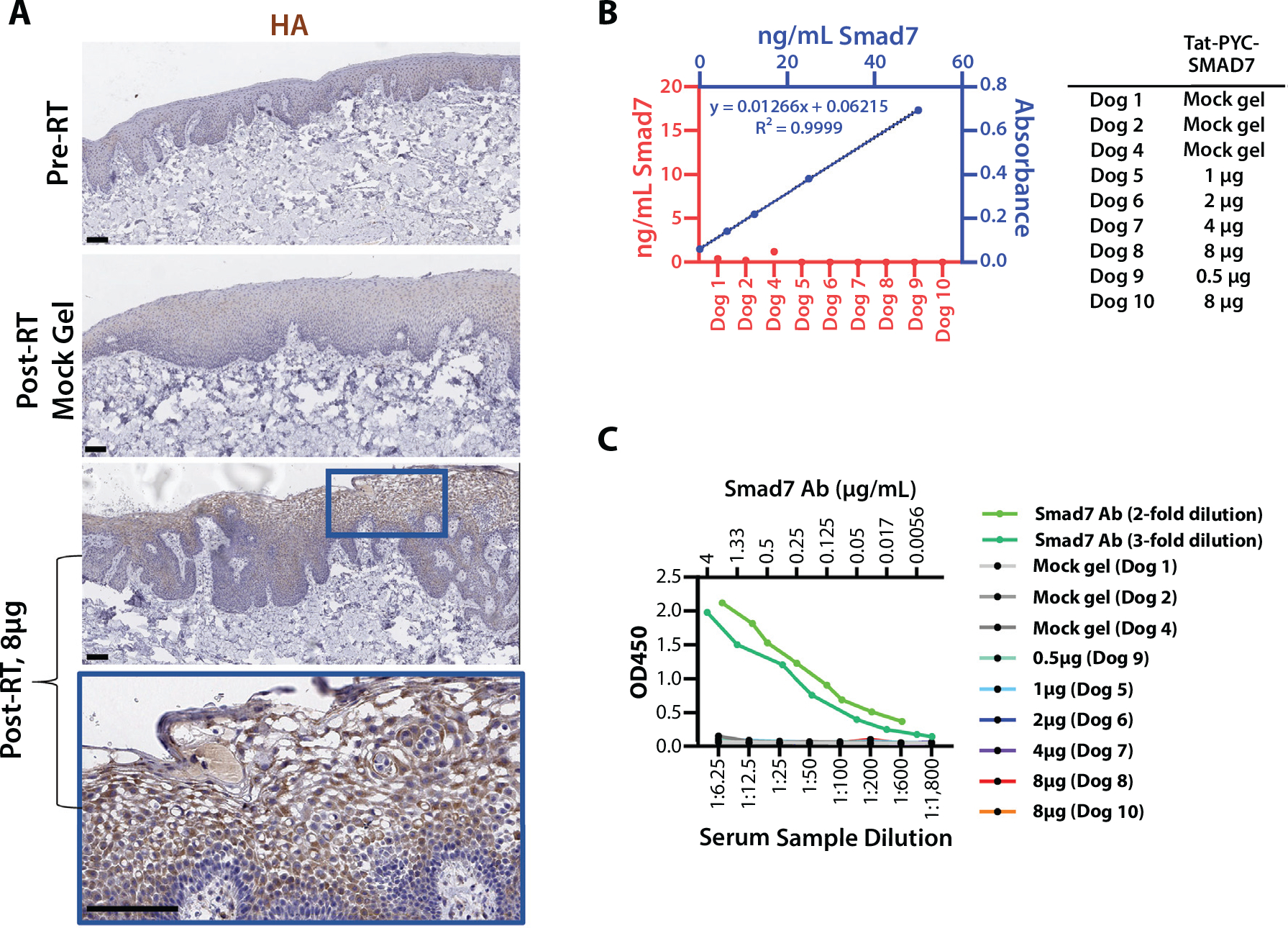
Fig. 3.

Tat-PYC-Smad7 targets local tissue. (A) Cell penetration of topical drug in RT irradiated oral mucosa. Antibody against c-terminal hemagglutinin tag was used for immunostaining. Lower panels present high-power view of the blue box identified in the panel above. Scale bar: 100 μm. (B) Tat-PYC-Smad7 was not detected in serum samples of topically treated dogs. The plasma of 2 untreated dogs diluted 1:4 was spiked with 0 to 50 ng/mL Tat-PYC-Smad7 protein to generate standard curves, which were averaged (presented in blue). The amount of Smad7 in the posttreatment plasma (diluted 1:4) was measured against the standard curves. (C) Lack of anti-drug antibody (ADA) against Tat-PYC-Smad7 from locally treated oral mucositis. Two independent assays (2 standard curves in light and dark green) for ADA examinations. No ADA against Tat-PYC-Smad7 was detected in dogs with oral mucosa Tat-PYC-Smad7 treatment for 2 weeks. Abbreviation: RT = radiation therapy.
