
Fig. 5.
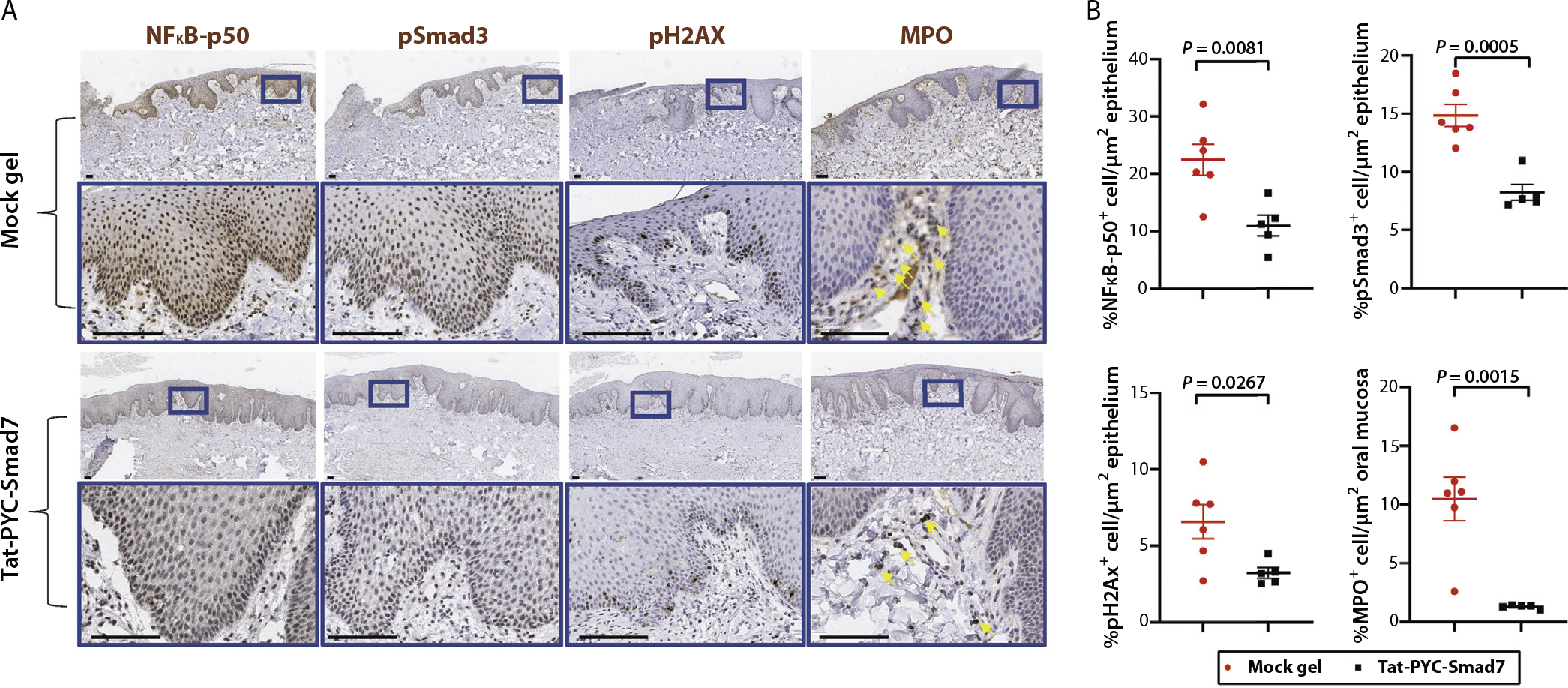
Pharmacodynamics markers of Tat-PYC-Smad7 treated dog oral mucositis. (A) Biopsies of posttreatment oral mucosa were evaluated by immunohistochemistry to detect NFkB-p50, pSmad3, pH2AX, and myeloperoxidase. Representative images of vehicle treated and Tat-PYC-Smad7 treated mucosa are presented with the lower panel in each set, presenting a high-power view of the blue box identified in the panel above. Scale bar: 100 μm. (B) Quantification of positive, brown-staining in epithelium sections excluding ulcers (in vehicle group) pictured in panel A. Myeloperoxidase staining together with morphology evaluation were used to identify neutrophils (indicated by yellow arrows) in ulcer surface and stroma. Faint brown staining in epithelium is nonspecific. The staining quantification for each dog and the mean of each treatment ± standard error of the mean is presented.