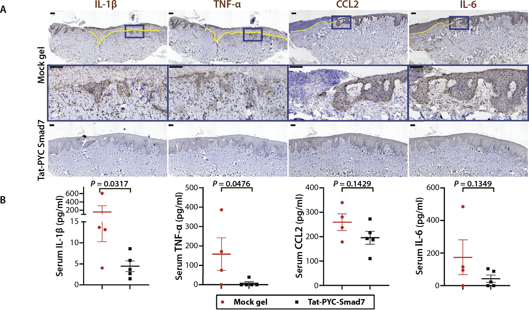
Fig. 6.

Tat-PYC-Smad7 reduced inflammatory cytokines released from oral mucositis lesions. (A) Immunohistochemistry showing inflammatory cytokines detected in mucosal epithelial and stromal cells. Yellow line highlights the ulcerated region. Insets (blue boxes) on top panels of vehicle group are enlarged in the panels below. Scale bar is 100 μm. (B) Serum detection of cytokines shown in A. Vehicle: mock gel from 4 dog biopsies with vehicle treatment only after radiation therapy; Tat-PYC-Smad7: from 5 dog biopsies treated with 1, 2, 4, and 8 μg daily dose of Tat-PYC-Smad7 treatment after radiation therapy. The serum quantification for each dog and the mean of each treatment ± standard error of the mean is presented.
