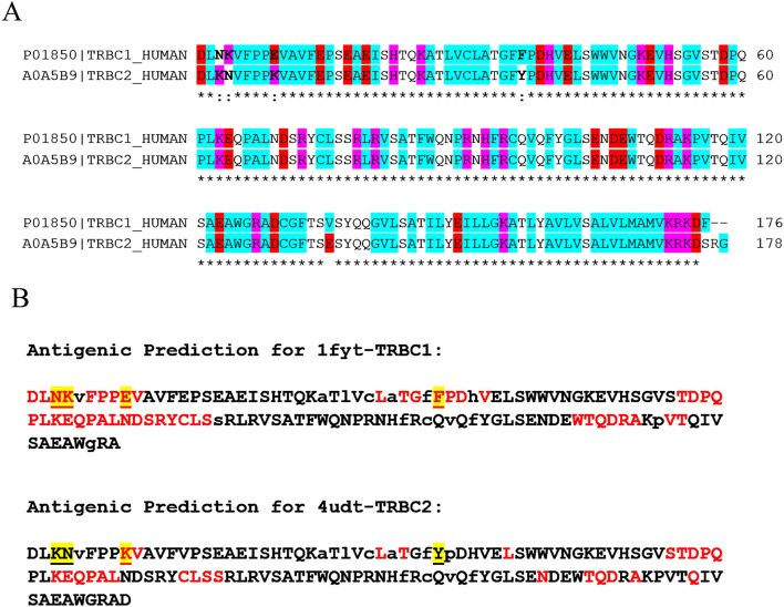
Figure 1.
Sequence alignment and antigenic prediction of TRBC proteins. (A) Sequence alignment of both TRBC proteins. TRBC1 and TRBC2 sequences were taken from Uniprot ID P01850 and A0A5B9, respectively. The red, pink and light blue highlighted negatively charged, positively charged and non-polar amino acids. The asterisk (*) denoted the identical amino acid residues between TRBC proteins. (B) Antigenic prediction of both TRBC proteins. TRBC1 and TRBC2 sequences were taken from the crystal structure PDB code 1fyt and 4udt, respectively. The capital and lowercase letters indicated surface and buried residues. The red letter indicated epitope residues. The highlighted and underlined letters indicate unique residues.

