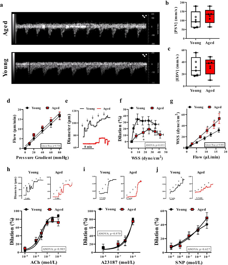
Fig. 1.
Impaired flow/WSS-induced dilation in skeletal muscle arteries of aged mice. Representative images (a) and summary data of peak systolic velocity (PSV, b) and end diastolic velocity (EDV, c) measured by Doppler ultrasound in the femoral artery from aged and young mice (n = 5 in each group). d–g. Stepwise increases in the pressure gradient induced a similar increase in intraluminal flow of isolated skeletal (m. gracilis) arteries in aged and young mice (d). Representative original traces (e) and summary data (f) of percent changes in diameter of arteries in response to increased WSS in young vs. age mice. Flow–WSS relationship is shown on Panel g. Representative original traces and summary data of percent diameter changes of isolated arteries in response to cumulative concentrations of acetylcholine (ACh, 10−9 to 10−6 mol/L) (h), Ca2+ ionophore, A23187 (10−9 to 10−6 mol/L) (i) or NO donor, sodium nitroprusside (SNP, 10−9 to 10−5 mol/L) (j) in young and aged mice. Arrows indicate stepwise increasing of flow or administration of agonists. Data are mean ± SEM or presented as box-and-whisker plots, in which the minimum, the 25th percentile, the median, the 75th percentile, and the maximum are shown, + indicates the mean of values. For statistical analyses of data on Student’s t test, repeated measures two-way ANOVA (insets: ANOVA p value for age effect is shown) with Sidak multiple comparisons were used. *p < 0.05 young vs. aged

