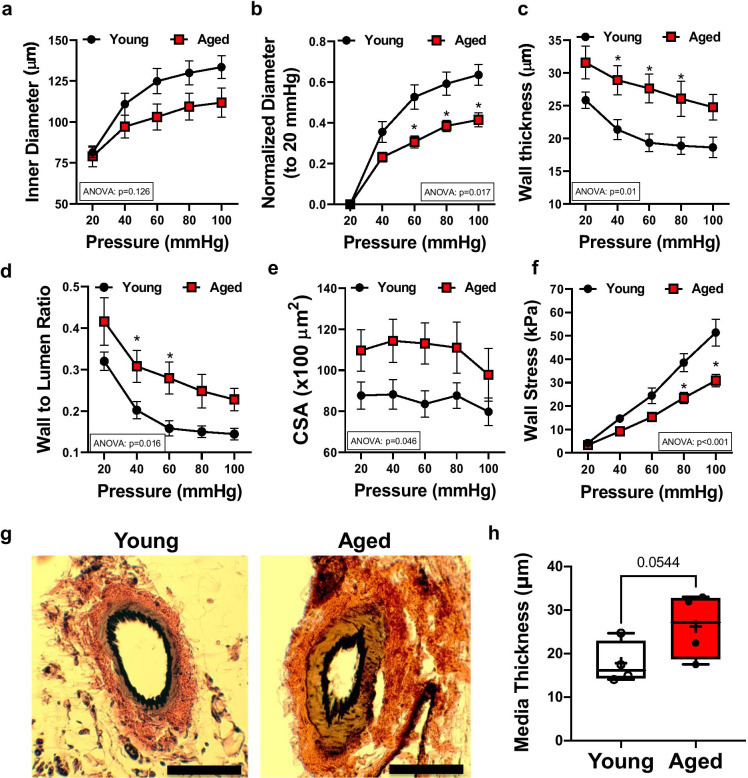
Fig. 2.
Hypertrophic inward remodeling in skeletal muscle arteries of aged mice. Skeletal muscle (m. gracilis) artery lumen diameter and vascular wall characteristic in response to increases in intraluminal pressure, ex vivo. Inner diameter (a), normalized diameter (to initial diameter at 20 mmHg) (b), wall thickness (c), wall-to-lumen ratio (d), cross-sectional area (CSA) (e), and calculated wall stress (f) in relation to increases in intraluminal pressure from 20 to 100 mmHg in skeletal muscle arteries isolated from young (n = 9) and aged (n = 6) mice. Data are mean ± SEM. For data analyses on panelse (a–h), repeated measures two-way ANOVA (insets: ANOVA p value for age effect is shown) with Sidak multiple comparisons were used. *p < 0.05 young vs. aged. Representative microphotographs of Elastin Verhoeff Van Gieson staining (EVG, elastic fibers and nuclei being stained black, collagen stained red, and cytoplasmic elements stained yellow/brown, scale bar: 50 μm) (g). Media thickness of arteries was measured by an unbiased orthogonal intercept method (Stereo Investigator, MBF) (h). Data are presented as box-and-whisker plots, in which the minimum, the 25th percentile, the median, the 75th percentile, and the maximum are shown, + indicates the mean of values. For statistical analyses of data on panel h, Studentis t test was used

