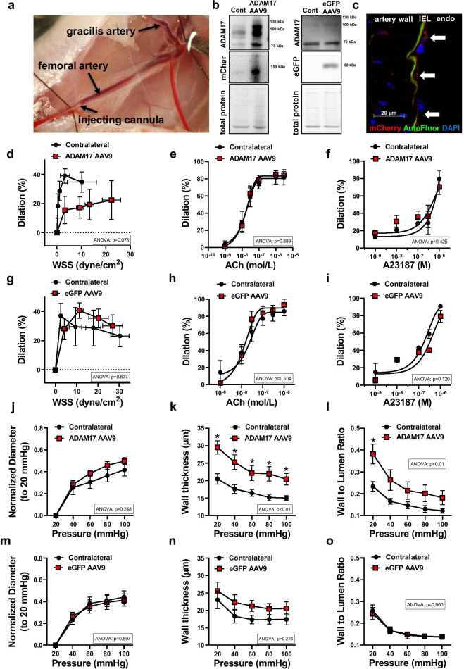
Fig. 4.
Impaired WSS mechanosensing and vascular remodeling after in vivo overexpression of ADAM17 in arteries of young mice. Representative photo depicts a procedure of intraarterial microinjection of ADAM17 encoded adeno-associated virus 9 (AAV9, labeled with mCherry) into the left skeletal muscle artery of the young mice (a). Representative images of Western immunoblot for detecting protein expression of ADAM17, mCherry, or eGFP in left (ipsilateral injected) and contralateral (Cont) skeletal muscle arteries are shown (b). Immunofluorescent staining for mCherry (shown in red, white arrow) was employed to confirm endothelial expression of the exogenous, AAV9 delivered ADAM17 in the left (ipsilateral) skeletal muscle arterial endothelium (endo). Interna elastic lamina (IEL) is shown in green with autofluorescence (AutoFluor). Nuclei were stained with DAPI (blue) (c). Summary data (n = 6–8, in each group) of percent diameter changes of isolated left ipsilateral (ADAM17 AAV9 or eGFP AAV9 injected) and right (contralateral) skeletal (m. gracilis) arteries in response to increased flow/WSS (d, g) or cumulative concentrations of acetylcholine (ACh, 10−9 to 10−6 mol/L) (e, h) and Ca2+ ionophore, A23187 (10−9 to 10−6 mol/L) (f, i). Summary data (n = 6–8, in each group) of inner normalized diameter (to initial diameter at 20 mmHg) (j, m), wall thickness (k, n), and wall-to-lumen ratio (l, o) in relation to increases in intraluminal pressure from 20 to 100 mmHg in isolated left ipsilateral (ADAM17 AAV9 or eGFP AAV9 injected) and right (contralateral) skeletal (m. gracilis) arteries. Data are mean ± SEM. For data analyses repeated measures two-way ANOVA (ANOVA p value for the corresponding effects are shown in the insets) with Sidak multiple comparisons were used. *p < 0.05 contralateral vs. ipsilateral artery

