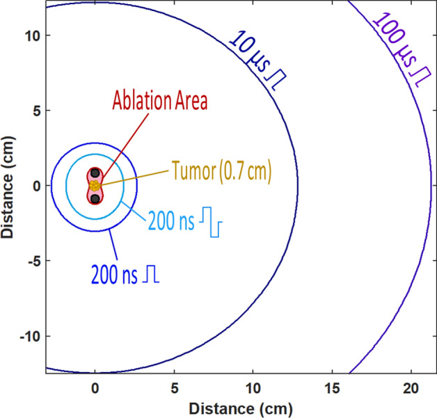
Figure 10.

The projected areas of nerve stimulation when a hypothetical tumor is ablated by pulses of different shape and duration. This example uses data from Figs. 2F and 9B and assumes uniform dielectric properties of the tissue. The tumor is placed between two needle electrodes which pierce the tissue orthogonal to the plane shown in the figure. Pulse amplitude is set to produce the electric field sufficient to ablate the tumor (“Ablation Area”). The larger radii mark the projected distances at which the nerve stimulation is expected when the tumor is ablated by pulses of a specific shape and duration (legends). See Figs. 2, 9, and text for more details.
