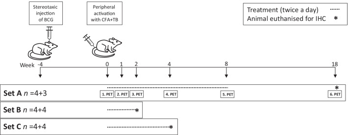
Fig. 1.
Study timeline. All animals (n = 24) were operated in week—4 and heat-killed bacillus Calmette–Guérin (BCG, 105 organisms in 2 μL of phosphate-buffered saline) was injected stereotaxically in the right striatum over 10 min. At week 0, the animals were intradermally injected with heat-killed mycobacterium tuberculosis (TB; 1,5 mg) in complete Freund’s adjuvant (CFA) to activate the brain lesion peripherally. Dosing of dimethyl fumarate (DMF; 15 mg/kg) or vehicle (tap water with 0.08% Methocel) using oral gavage was started on day 0 in week 0. Set A animals had a baseline PET study on the same day. Set A animals were imaged during the treatment regimen in weeks 1, 2, 4, and 8, as well as 10 weeks after the treatment finished (i.e. week 18). One Set A animal from the control group died after imaging in week 4. Set B animals were euthanised for immunohistochemistry (IHC) in week 2 and set C animals were euthanised for IHC in week 4

