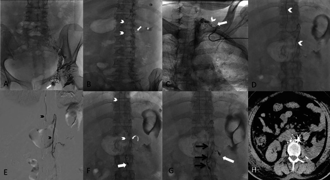
Fig. 1.
A Post intranodal lymphangiography from left inguinal node (white arrow), shows gradual opacification of lymphatic channels cranially with lipiodol reaching upto the lower abdomen. B The lumbar lymphatic duct (arrowheads) is visualized along with the site of leak (arrow) and the extravasation (asterisk). C Using retrograde left subclavian approach, the venolymphatic junction is cannulated (arrow) and microcatheter is progressed into thoracic duct (arrowhead). D Microcatheter (arrowheads) is advanced upto the leak site by selectively cannulating the left lumbar lymphatic branch. E A ductogram is taken with iodinated contrast demonstrating the lymphatic duct (black arrowheads) and extravasation in the left renal fossa. F Microcatheter (arrowheads) is advanced beyond the site of leak (curved arrow) and coil embolization done (white arrow). G Glue embolization done with 33% N-butyl cyanoacrylate and the glue cast is appreciated in the lymphatic duct (black arrows) and extending into the site of leak (white arrow). H Axial computed tomography image at mid abdomen level, the next day demonstrating post embolization glue accumulation with coils at the leakage site, showing steak artifact with minimal residual ascites

