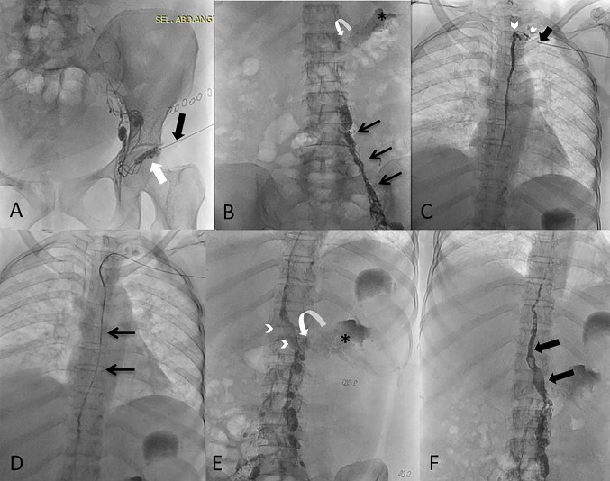
Fig. 2.
A Intranodal lymphangiography from left inguinal node (white arrow) with 25 G needle (black arrow). B Figure shows gradual opacification of lymphatic channels cranially (black arrows) with lipiodol reaching upto the lower abdomen. The site of leak (curved arrow) is visualized along with and the extravasation (asterisk). C Using retrograde left subclavian approach, the venolymphatic junction is cannulated (black arrow) and microcatheter is progressed into thoracic duct (arrowheads) and contrast was injected into thoracic duct. D Microcatheter (arrows) is advanced beyond into the abdominal part of thoracic duct. E Glue embolization done with N-butyl cyanoacrylate in the selective lymphatic duct (arrowheads) and extending into the site of leak (curved arrow) with visualization of site of leak (asterisk). F Post embolization glue cast (black arrows) can be appreciated in the lymphatic, however no active chylous leak was noted in the left renal fossa

