Figure 3.
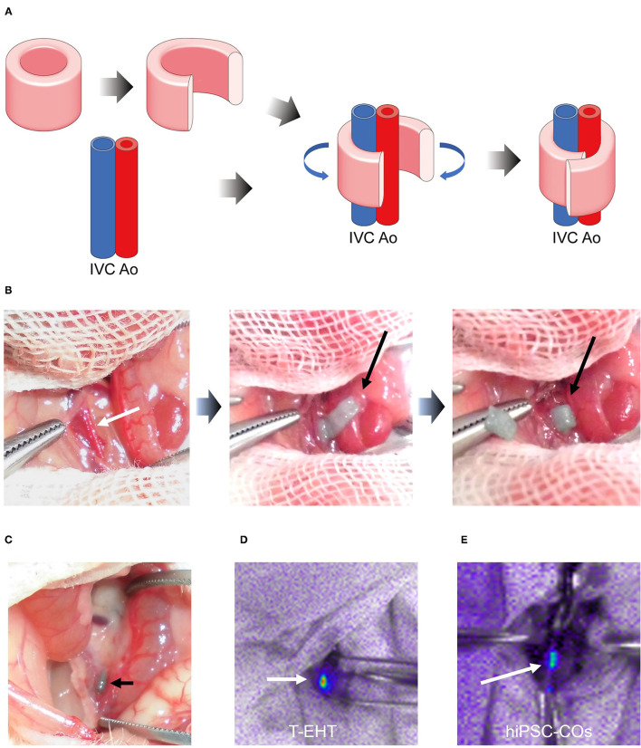
Transplantation procedure and histological analysis of transplanted tubular engineered heart tissue (T-EHT). (A) Scheme of transplantation procedure. The T-EHT is removed from the needle array, cut open, and wrapped around the abdominal aorta and inferior vena cava (IVC). (B) Transplantation procedure. The abdomen of each NOG mouse is opened under general anesthesia. The aorta and IVC are exposed (white arrow) and the T-EHT (black arrow) is wrapped around them. (C) After 1 w of transplantation. T-EHT (black arrow) is engrafted in the NOG mice. (D) and (E) Fluorescent images captured after 1 w of transplantation. The T-EHT and hiPSC-COs were labeled with far-red before transplantation (white arrows). Engrafted tissues with far-red fluorescence were visualized using Nightowl.

