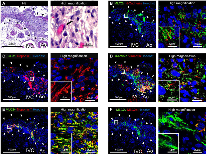
Figure 4.
Histological staining of the tubular engineered heart tissues (T-EHTs) and its high magnifications images after 1 w of transplantation. (A) Hematoxylin and eosin (HE) staining. (B) Myosin light chain 2v (MLC2v) and N-cadherin. (C) CD31 and cardiac troponin T (cTnT). (D) α-actinin and vimentin. (E) MLC2v and cTnT. (F) MLC2v and myosin light chain 2a (MLC2a). The HE staining shows engraftment of the T-EHTs (black arrowheads) (A). Red blood cells were found in the luminal structure with endothelial cells (black arrow). Immune staining reveals MLC2v-positive cells and N-cadherin-positive cells between them (B). The cTnT-positive cells are found in the peripheral area of the tissue, and vimentin-positive cells are found in the middle (C,D). Muscle striations are observed in MLC2a, MLC2v, and α-actinin staining (D–F).

