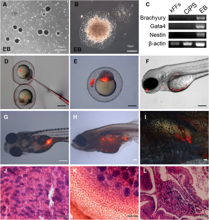
FIGURE 3.
Differentiation potential of Kio-CiPSLCs derived from kFFs. (A,B) The embryoid body (EB) formation (A) and differentiation (B). (C) RT-PCR analysis of ectoderm marker gene (Nestin), mesoderm marker gene (Brachyury), and endoderm marker gene (Gata4) in the three germ layers. (D–F) Different distributions of PKH26-labeled donor cells (Kio-CiPSLCs, passage 15, red) in different stages, in which the chimeras were analyzed by microscopy at 5 days post-fertilization (F). Scale bars, 200 μm. (G–I) PKH26-labeled Kio-CiPSLCs (passage 15, red) were transplanted with approximately 1,000 cells to host zebrafish, and the teratomas were analyzed after 8 weeks post-injection. Of the 30 injected fish, teratomas were observed in five fish. Scale bars, 200 μm. (J–L) Histology of various tissues present in teratomas derived from Kio-CiPSLCs. Neural epithelium (ectoderm) (J); Cartilage (mesoderm) (K); Gut-like epithelium (endoderm) (L). Scale bars, 50 μm.

