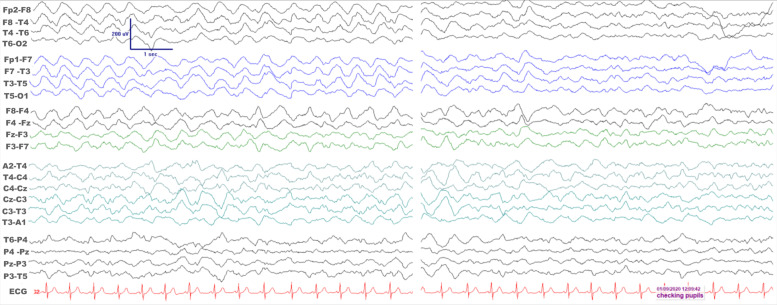
Figure 1.
Electroencephalogram (EEG) recordings in the intensive care unit while the patient was comatose. A bipolar montage is shown while the recording was undertaken according to the 10–20 international system of electrode placement. There is remarkable bilateral synchrony of anteriorly maximal rather monorhythmic high amplitude delta frequency activity on the left EEG epoch. On the right, there is significant EEG reactivity as the patient’s pupils were examined.

