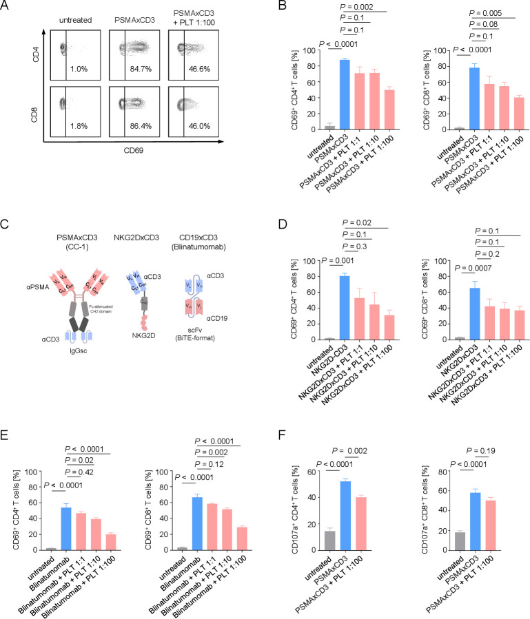
Figure 2.
Platelets impair the therapeutic T-cell activation capacity of bispecific antibodies. (A, B) Activation of CD4+ and CD8+ T cells was determined by analysis of CD69 expression after 24 hours. PBMCs of healthy donors were incubated with LNCaP cells (E:T 4:1) in the presence or absence of PSMA×CD3 (200 ng/mL) and with the indicated E:P ratios. (A) Exemplary flow cytometry results and (B) combined data are shown (n=3). (C) Schematic illustration of the different bispecific T cell-engaging formats used in our study. (D, E) Activation of CD4+ and CD8+ T cells was analyzed on coculture of PBMCs of healthy donors with target cells at an E:T ratio of 2.5:1.0 and the indicated E:P ratios for 24 hours. (D) The sarcoma cell line SaOS-2 was cultured with PBMCs (n=3) and NKG2DxCD3 (2.5 µg/mL). (E) The B-ALL cell line (SEM) was cultured with PBMCs (n=4) and CD19xCD3 (blinatumomab, 200 pg/mL). (F) Degranulation of CD4+ and CD8+ T cells was determined by analysis of CD107a expression after 4 hours. PBMCs of healthy donors were cultivated with LNCaP cells at an E:T ratio of 4:1 in the presence or absence of PSMA×CD3 (200 ng/mL) or platelets (E:P 1:100). Pooled data (n=5) are shown. (B, D–F) Statistical significance was calculated by one-way analysis of variance and Tukey’s multiple comparisons test. E:P, effector to platelet; E:T, effector to target; PLT, platelets; PBMC, peripheral blood mononuclear cell.

