
Figure 1. The RASSF1A expression pattern is reciprocal to HES1 across human tumor types.
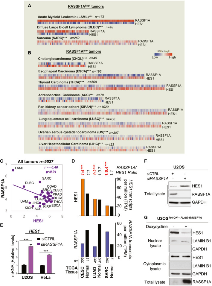
- Correlation heatmaps depicting an opposite RASSF1A‐HES1 expression pattern across the tumors with highest expression of RASSF1A. RNA seq data were retrieved from the TCGA database. Of the 32 tumor types of the TCGA database included in the analysis, tumor types with an average of > 4.2 (median) RASSF1A Transcripts per million (TPM) were classified as RASSF1Ahigh, whereas tumor types with an average of < 4.2 RASSF1A TPM were classified as RASSF1Alow. See also Dataset EV1.
- Same as A, for the TCGA tumors with the lowest expression of RASSF1A. See also Dataset EV1.
- RASSF1A (bottom) and HES1 (top) transcripts per million (TPM) in the indicated types of human tumors and respective normal tissue. Data were extracted from the TCGA and GTEx databases using the GEPIA/GEPIA2 online tool for normal and cancer gene expression profiling and interactive analyses. The number of patients is indicated in each tumor type. See also Dataset EV1.
- qPCR for HES1 mRNA levels in siCTRL (non‐targeting) and siRASSF1A‐transfected U2OS and HeLa cells.
- U2OS cells were transfected with either siCTRL or siRASSF1A and lysates were immunoblotted for HES1 levels.
- U2OS cells Tet‐On inducibly expressing FLAG‐RASSF1A were treated with doxycycline at a concentration of 0.5 μg/ml for 24 h. The cells were fractionated in order to acquire the nuclear and cytoplasmic extracts, which were subsequently Western blotted and probed with the indicated antibodies.
Data information: Tumor type abbreviations: ACC: Adrenocortical carcinoma; BLCA: Breast invasive carcinoma; BRCA: Breast invasive carcinoma; CESC: Cervical squamous cell carcinoma and endocervical adenocarcinoma; CHOL: Cholangiocarcinoma; COAD: Colon adenocarcinoma; DLBC: Lymphoid Neoplasm Diffuse Large B‐cell Lymphoma; ESCA: Esophageal carcinoma; KICH: Kidney Chromophobe; KIRC: Kidney renal clear cell carcinoma; KIRP: Kidney renal papillary cell carcinoma; KIPAN: Pan‐kidney cohort (KIRP + KIRC + KICH); LAML: Acute Myeloid Leukemia; LIHC: Liver hepatocellular carcinoma; LUSC: Lung squamous cell carcinoma; OV: Ovarian serous cystadenocarcinoma; PRAD: Prostate adenocarcinoma; SARC: Sarcoma; THCA: Thyroid carcinoma; UVM: Uveal melanoma. ***P < 0.001 of Student’s t‐test. Error bars indicate s.e.m. Data shown are representative of three biological replicates (n = 3).
Source data are available online for this figure.